A grating based interferometer X-ray phase-contrast (GIXF) setup (Figure 1) has been added at the existing Rigaku rotating anode at HCØ at University of Copenhagen. The setup is running at 50 kV and 200 mA. The first image of a Legoman is shown in Figure 2. Images and tomograms are now been routinely obtained, but there was a major breakdown of the anode just after the end of CIA-CT. However, the setup up runs again with a visibility of about 13 % in the fringes. This could probably be improved with new and optimized gratings. Currently the setup uses either to the 3rd or the 5th fractional Talbot distance for various applications. The application area is normally within food science although other sample types are also tested.
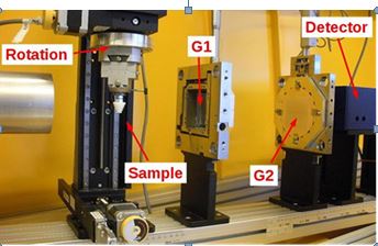
Figure 1 : X-ray setup at NBI. The gratings are to the right.
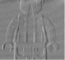
Figure 2: Phase contrast image of a LEGO man [Con4].
Both phase contrast and dark-field imaging have been implemented and the results have been obtained and are published in the journal Food Control [PJ11]. The focus so far has been on foreign body detection in food products where dark-field imaging shows great potential as a complementary imaging modality to conventional absorption contrast imaging. The first data set was part of Mikkel Schou Nielsen’s MSc thesis [MSc10] and showed the potential of the technique for foreign body detection. The second article shows the potential of the dark field technique for detecting frost damages in berries and fruit. Apparently frost damages in berries causes cell damages that are visible in dark field imaging, but not in normal absorption mode. This effect will be investigated further in the future and might be used for applications. In the spring of 2012, a project on using new X-ray imaging modalities, in particular absorption and dark-field imaging, for quality assessment of chocolate products was performed in collaboration with the company TOMs (see application example in Figure 3). Today, the quality assurance for monitoring of the correct filling in chocolate plates is cumbersome. The aim of the investigation was to see if X-ray imaging could be used to measure the content of filling in a chocolate plate.
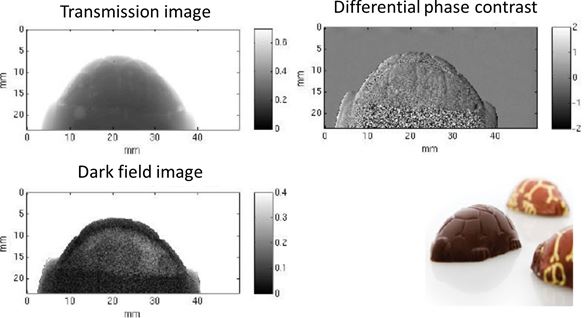
Figure 3: Radiograms of a "Toms skildpadde" chocolate with filling using absorption- (upper left), refraction- (upper right) and scattering-based contrast. The contrast between filling and chocolate is largest using the scattering contrast [New6].
Dark field imaging has also been tested in a metrological study in a collaboration between NBI and DTU and a publication has been accepted. A range of new applications of dark field imaging in materials science are being pursued. A paper has been accepted in Applied Physics A showing how dark field imaging can be used to map misorientations of fibers [PJ16]. This could be of interest in fiber enforced materials. The techniques can also be used for mapping of very small cracks that are invisible by absorption based methods when the cracks are of size below the pixel size of the tomograms. Two more articles using this approach are under preparation. Besides the laboratory work, analysis of X-ray data measured on a grating based set up at Technische Universität München (TUM) has been done. We measured on beef samples from DMRI. The original purpose was to investigate if it was possible to detect differences in beef tenderness from the dark field or electron density tomograms obtained by the setup. We have not yet been able to distinguish beef quality from our data, but the data analysis has led to the creation of 2D (electron density, absorption length) histograms (Figure 4) of the tomogram data. These tomograms represent an X-ray refractive index distribution, and can be expanded to 3D by incorporating the incoherent scattering length from the dark field tomograms. In total this 3D distribution gives a unique and new overview of the optical characteristics of a given sample. The method has been published in Phys.Med.Biol. (2012).
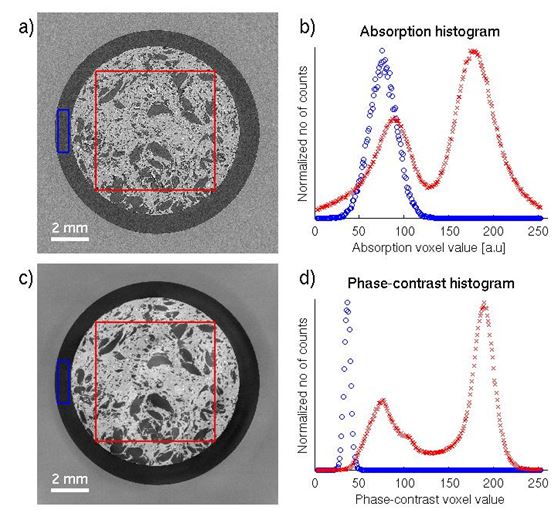
Figure 4 : Dual Histogram of absorption and phase contrast data.
In July of 2012 in collaboration with a research group at LIFE, CT-measurements on meat samples were performed at a synchrotron facility. The samples were emulsions of protein and fat used for sausages. The aim of the measurement was preliminary investigations of what information CT-measurements can give on heterogeneous meat products, and to evaluate if new imaging modalities can provide added contrast and better signal quality than conventional CT. As seen in Figure 5, preliminary studies show that there are clear improvement in SNR and CNR when using phase-contrast CT measurements for studying meat products.
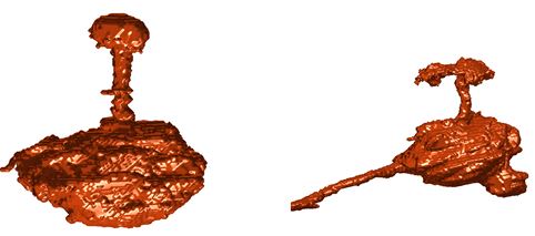
Figure 5: Absorption (top) and phase-contrast (bottom) CT measurements show that the phase-contrast CT has a superior SNR and CNR between the fat and protein. The blue graph in the histograms (right) which show the distribution of voxel values of the Eppendorf tube and the red graph of the meat sample confirm this.
Work is currently including development of new methods for image processing of meat samples and two publications are under preparation. Experiments with Insulin injections in pork meat simulation in injections in human tissue have also been performed in collaboration with Novo Nordisk. The aim is to investigate the shape of the insulin depot after injection and how it influences the effect of the injection. Measurements have mostly been performed in absorption mode using Iodine as a contrast agent. Examples are shown in Figure 6.
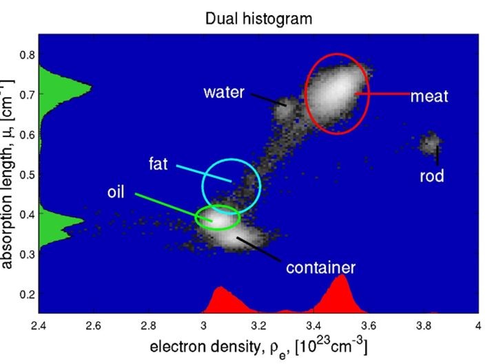
Figure 6: Two different insulin depots after injection in pork meat [MSc8].
Experiments have been performed on pork fat and published in Meat science. Phase contrast measurements give a significant better contrast. Enhancing between meat and cartilage the contrast sufficiently to discriminate the two has not yet been successful. In collaboration with the Department of Oral & Maxillofacial Surgery, Institute of Odontology, Faculty of Health Sciences, University of Copenhagen a novel approach to analysis of bone integration for dental implants has been tested. The method uses 3D tomograms rather than the 2D cross sections investigated using light microscopes that are traditionally used for evaluation. The method gives an overall picture of the radial dependencies of some key parameters (bone fraction, bone density) for the whole volume surrounding the implant. A paper is being prepared for submission. The CIA-CT project has given us a perfect platform to develop the grating based technique and apply for new funding. Funding from the strategic research council has been granted for a project entitled NEXIM: New X-ray Imaging Modalities - For Safe and High Quality Food. The project is a collaboration between the Niels Bohr Institute and the Department of Food Science at KU, Danish Meat Research Institute at TI and Department of Informatics and Mathematical Modelling at DTU. We have also made close contacts with a range of companies and Institutes that we are now collaborating with. Furthermore we are participating in the p3 project funded by Højteknologifonden and the CINeMA project funded by the strategic research council.